
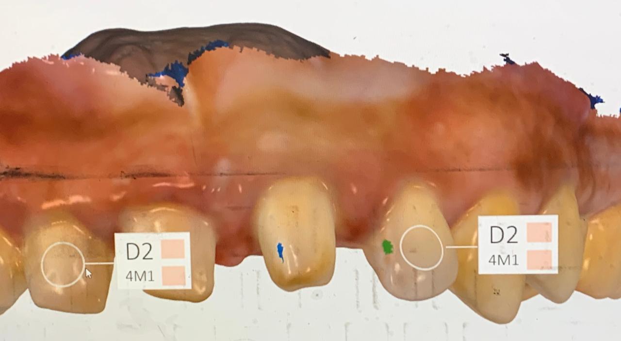
Understanding Crown Margins: A Deep Dive into Design, Materials, and Clinical Success through Digital Integration
Dr. Syed Nabeel, BDS, D.Orth, MFD RCS (Ireland), MFDS RCPS (Glasgow)
Dr. Syed Nabeel – Dedicated to Neuromuscular Dentistry, Orthodontics & Digital Innovation
Dr. Syed Nabeel is a dentist and innovator committed to education, patient care, orthodontics, and research. As Founder & CEO of DentistryUnited.com (since 2004), he has built a global platform for dental professionals. He also launched Dental Follicle – The E-Journal of Dentistry (ISSN 2230-9489) in 2006 to promote scholarly exchange in dentistry and orthodontics.
Clinical Practice & Leadership
As Managing Director of Smile Maker Clinics Pvt Ltd, ( www.smilemaker.in ) Dr. Nabeel manages his practices and plans nationwide expansion with a focus on evidence-based dentistry and research. His areas of focus include:
✔ Neuromuscular Dentistry & TMJ Treatment
✔ Full-Mouth Rehabilitation & Smile Makeovers
✔ Orthodontics – Braces, Aligners & Digital Treatment Planning
25 Years of Learning & Innovation in Dentistry & Orthodontics
With 25 years of experience, Dr. Nabeel continues to explore AI, orthodontics, and digital workflows to enhance patient care. His key interests include:
AI in Dentistry & Orthodontics – Improving diagnostics & treatment precision
Digital Dentistry & Workflow Optimization – Enhancing efficiency & patient experience
Educator & Speaker
A dedicated mentor & speaker, he enjoys sharing insights on:
✔ Neuromuscular Dentistry, Orthodontics & TMJ Disorders
✔ Practice Management & Digital Integration
Beyond Dentistry
Dr. Nabeel finds joy in wildlife photography, travel, and gardening, always eager to learn from new experiences. Grateful for his mentors, colleagues, and patients, he remains committed to growth and innovation in dentistry and orthodontics.
Email: dentistryunited@gmail.com
Website: www.DentistryUnited.com