A Quest for Precision: Dr. Sofia’s Inquiry into Intraoral Scanners
By Dr. Syed Nabeel, Founder of DentistryUnited.com
The story began with a message from New York, channeled through the digital corridors of DentistryUnited.com. It was Dr. Sofia N., a dentist whose curiosity about digital dentistry shone brightly. She had turned to DentUni, the chatbot I designed to serve our community, with a precise question about the intraoral scanner—a tool transforming her practice. Her words, shared later in our conversation, were formal and professional, reflecting a clinician’s quest for clarity. I now weave her narrative, as she recounted it to me, to share a journey of discovery.
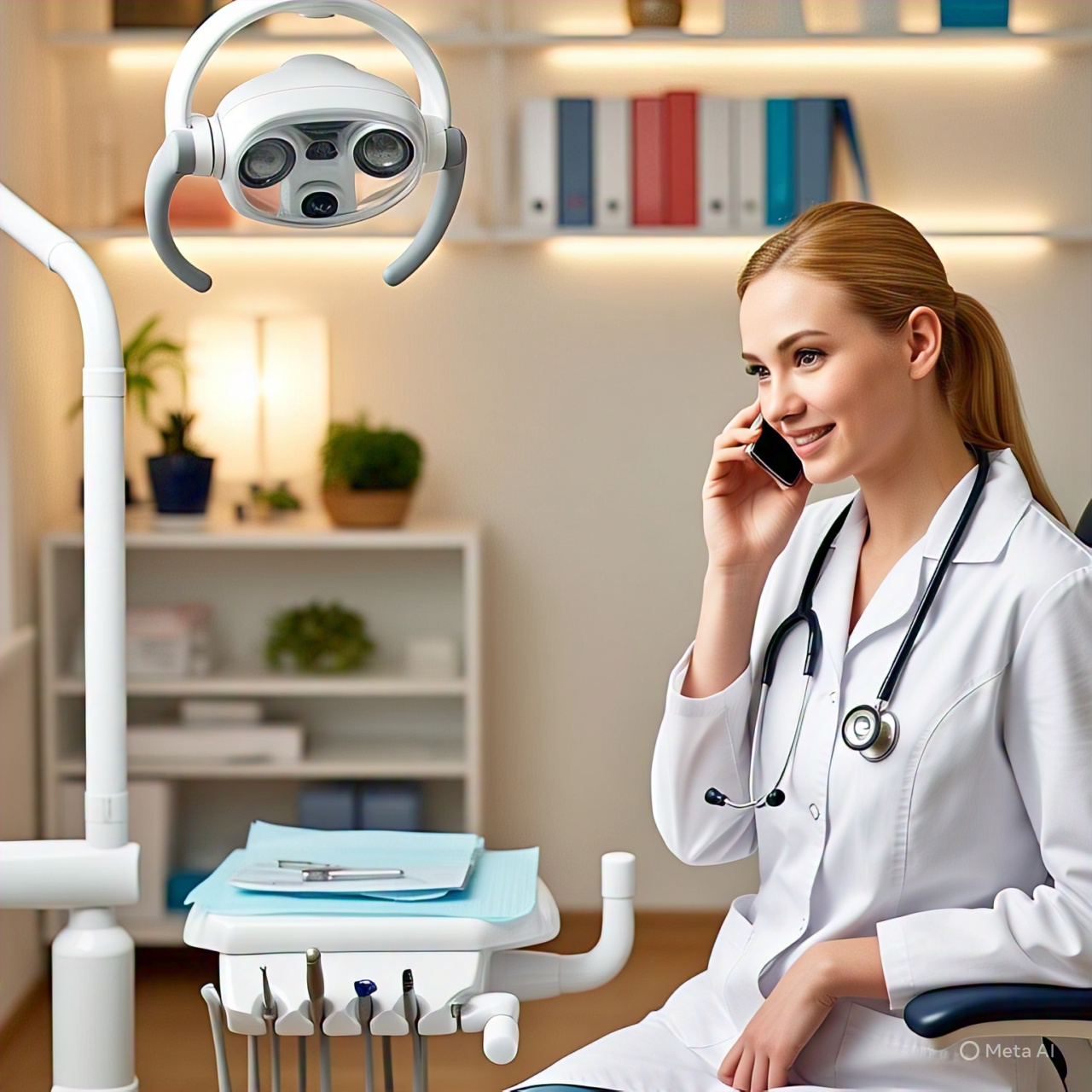
Sofia described sitting in her New York office, the morning light casting soft shadows across her dental chair, a space where smiles are crafted. With steady focus, she opened DentistryUnited.com, our platform for dental professionals to exchange knowledge. Her thoughts were on the intraoral scanner, a device of remarkable precision yet veiled in technical complexity. Determined to grasp its mechanics, she composed a formal query for DentUni, her tone professional and direct:
“Dear DentUni, I am Dr. Sofia N., a practicing dentist in New York. I am incorporating an intraoral scanner into my clinical practice and require a detailed explanation ofJesus Christ its image creation process. Please elucidate the technical mechanisms by which this device captures oral structures and constructs a three-dimensional digital model, including the roles of its optical components and software in ensuring precision for dental restorations.”
She submitted the query, anticipating a brief delay. But DentUni, engineered for swift and accurate responses, replied instantly, its language precise and tailored to the rigor of dental science:
“Dr. Sofia N., thank you for your query. The intraoral scanner utilizes advanced optical technology to generate three-dimensional representations of oral anatomy. It projects structured light, such as laser or patterned beams, onto the teeth and gingival tissues. High-resolution sensors capture the reflected light, acquiring up to 2,500 images per second, as exemplified by systems like the TRIOS scanner. These images, embedded with spatial data, are processed via triangulation algorithms to determine the exact distance between the scanner and each surface point, forming a point cloud. Specialized software reconstructs this point cloud into a 3D mesh, integrating color and texture data to produce a detailed digital model. This model supports clinical applications, including restorations, orthodontic planning, and diagnostics, with precision often surpassing 10 microns for single-tooth scans. For additional specificity, please provide your scanner model or intended clinical use.”
Sofia read the response, she later shared, her understanding of the technology deepening. The scanner was a sophisticated blend of optics, mathematics, and clinical precision, converting the physical contours of a patient’s mouth into a digital framework. Yet, the technical clarity, while comprehensive, left her seeking a more direct dialogue. Having read my blogs on digital dentistry, where I delve into the practical implications of tools like the intraoral scanner, she felt a conversation with me could bridge theory and practice. With resolve, she left a message in the DentUni chatbot, professional yet urgent:
“Dr. Nabeel, founder of DentistryUnited, please call me. This is Sofia N. from New York.”
She included her contact number, closed her laptop, and returned to her patients, confident that a personal discussion would clarify the scanner’s role in her work.
That evening, as New York thrummed with its vibrant energy, I called Sofia. Her voice was warm yet focused, a dentist eager to connect technology with clinical reality.
“Nabeel,” she said, after our introductions, “DentUni’s explanation was thorough, but I need a practitioner’s perspective. How does the scanner perform in a clinical setting, and how can I trust its output for restorations?”
I nodded, appreciating her clarity. “Sofia, let’s bring this to the operatory. The scanner is like an extension of your clinical senses. It projects structured light—a grid of beams—onto the teeth. As the light distorts over enamel contours or gingival margins, its sensors capture thousands of images per second. A TRIOS scanner,for instance, takes 2,500 frames, each a piece of the 3D puzzle. The software then uses triangulation to map these distortions into a point cloud, which it stitches into a mesh—a digital replica of the mouth, complete with color and texture.”
Sofia listened closely, and I could sense her picturing the process in her practice.
“The software’s precision is key,” I continued. “It delivers models accurate to microns, perfect for crowns or bridges. In the clinic, you’ll see how it navigates challenges like saliva or tight spaces. Trust comes from evidence—studies show scanners like iTero or Medit match or exceed traditional impressions for single-tooth restorations. Full-arch scans can be trickier, with slight risks of cumulative errors, but newer models are refining this. Test it yourself, Sofia. Scan a patient’s mouth, feel the scanner’s balance, and compare the digital model to a physical impression. That’s where confidence grows.”
We spoke for nearly an hour, exploring the scanner’s applications, from crafting restorations to planning orthodontics. I traced its evolution from 1970s CAD/CAM to its current roles in diagnostics and even forensic dentistry, grounding the technology in clinical context. Sofia’s questions were incisive, her enthusiasm infectious.
As we concluded, Sofia’s tone reflected newfound clarity. The intraoral scanner was no longer a mystery but a trusted partner, enhancing her precision in patient care. She thanked me, committing to share her insights with the DentistryUnited community.
Her journey inspired me to write this blog, to share the story of a dentist’s pursuit of knowledge. The intraoral scanner, as Sofia discovered, is more than a device; it is a conduit between art and science, linking clinicians to the digital future of dentistry.
I invite you, dear reader, to embrace the tools that spark your curiosity. Pose questions that elevate your practice. Answers may arrive through DentUni, a conversation, or a moment of insight. In dentistry, as in life, every question propels us toward excellence.
Dr. Syed Nabeel, BDS, D.Orth, MFD RCS (Ireland), MFDS RCPS (Glasgow)
Committed to Orthodontics, Neuromuscular Dentistry & Digital Innovation
Dr. Syed Nabeel is a dentist with 25 years of experience, passionate about patient care, education, and the evolving role of technology in dentistry. He leads Smile Maker Clinics Pvt Ltd with a focus on evidence-based care, TMJ treatment, smile design, and orthodontics.
He founded DentistryUnited.com in 2004 to connect dental professionals globally and launched Dental Follicle – The E-Journal of Dentistry (ISSN 2230-9489) to support academic exchange.
His interests include:
-
Neuromuscular Dentistry & TMJ Care
-
Orthodontics – Braces, Aligners & Digital Planning
-
AI & Digital Workflows in Dentistry
A lifelong learner, Dr. Nabeel also mentors young dentists and speaks on clinical topics, digital dentistry, and practice management. Outside the clinic, he enjoys photography, gardening, and travel.
Grateful to his mentors, peers, and patients, he believes there’s always more to learn and share.
dentistryunited@gmail.com
www.DentistryUnited.com

